Sestamibi Scans and Ultrasound Scans.
Sestamibi and ultrasound scans are used to plan surgery in primary parathyroid disease.
These scans are not to be used for diagnosis - except under very rare and specific circumstances by the surgical team.
These scans are not to be used for diagnosis - except under very rare and specific circumstances by the surgical team.
Scans should not be used to determine who should have, or who should not have parathyroid surgery. Parathyroid scans will often fail to identify the usually single offending gland - so what - well if you are with an experienced parathyroid surgeon this will not put them off and indeed cure rates should still be excellent.
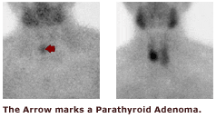
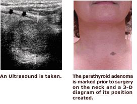
I do use scans to help plan surgery and indeed to hopefully facilitate minimal access parathyroid surgery. The scan images below show how in the top images a parathyroid adenoma can be seen on Sestamibi scanning and in the bottom image the ultrasound shows the 3D appearance.
Scan quality is crucial. I have seen a number of scans from hospitals which undertake small volumes of parathyroid imaging where the scans are of poor quality. This is not helpful as parathyroid adenomas may be missed on poor quality scans. Ideally all scans would be undertaken in centres where large numbers of scans are done each year and the surgeon and endocrinology team jointly review cases to discuss findings with the radiologists. This type of MDT (multi-disciplinary) team approach has been shown in many fields to improve outcomes.
No comments:
Post a Comment